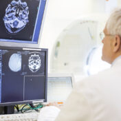
A CT scan (also known as a CAT scan) creates 3-D pictures of structures of the body using multiple low-intensity X-ray images with a computer that generates cross-sectional views (or “tomograms”). Unlike conventional x-ray images which shows only bone, CT images also show bones, blood vessels, muscles and organs. CT scans can also be used to assist in procedures by helping to guide the placement of instruments.
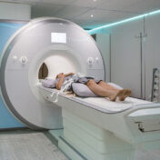
Magnetic Resonance Imaging (MRI) – Body
 moreless
An MRI machine uses a large magnet and radio waves to produce 3-D images of the body that can be viewed from many different angles. These highly detailed, cross-sectional images of the organs, tissues and bones of the body enables the discovery of abnormalities that might be obscured by bone with other imaging methods. Unlike x-rays and CT scans, an MRI scan does not use ionizing radiation.
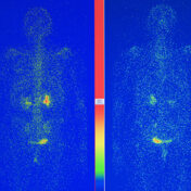
Nuclear Medicine
 moreless
A small amount of radio-active substance (a “tracer”) is injected into the patient which is then detected by a special camera in order to capture precise images of organs and tissues. Unlike other imaging modalities, nuclear imaging techniques show the physiological function of the tissue or organ being investigated, while other scans such as MRI and CT only show the anatomy. Nuclear Medicine can also be used in therapeutic procedures that are able to identify a disease in its earliest stages.
Positron Emission Tomography (PET)
 moreless
Positron emission tomography, also called a PET scan, is a type of nuclear medicine imaging that measures important body functions, such as oxygen use, blood flow, and glucose metabolism, which helps doctors determine how well organs and tissues are functioning. Unlike MRIs or CT scans, PET scans show metabolic changes occurring early on at the cellular level in an organ or tissue. This is important because disease often begins at the cellular level.
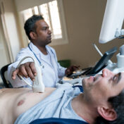
Ultrasound
 moreless
Ultrasound scans, or sonography, use high-frequency sound waves to create images of the inside of the body. Ultrasounds are safe because they use sound waves or echoes to create an image, instead of radiation. Varying shades of gray on the scan reflect different densities. It can be used to examine internal organs such as the liver and kidneys, pancreas, thyroid gland, testes and ovaries. It is also is suitable for use during pregnancy. In addition to being used for making diagnoses, ultrasounds are also valuable in guiding doctors with procedures such as biopsies.
Radiography (x-ray)
 moreless
Radiography, or an x-ray, is the oldest, most basic, and cost-effective form of medical imaging. A very small dose of radiation is passed through the body to create fixed images of the internal structures of the body. X-rays excel in detecting fractured bones, joint injuries, infections and foreign objects.